

ILD CHEST Capsule 2026 is a focused scientific initiative designed to enhance clinical expertise in interstitial lung diseases through structured, case-based learning. Featuring real-world scenarios with multidisciplinary perspectives from leading Indian pulmonologists, radiologists, pathologists and rheumatologists, the program supports accurate diagnosis, strengthens clinical decision-making, and promotes evidence-based management in practice.
A 66-year-old woman, a never-smoker, presented with progressive exertional dyspnea, chronic nonproductive cough, fatigue, and a 15-lb unintentional weight loss over 6 months. She reported avid gardening and long-term exposure to a pet cockatiel. Examination revealed subtle inspiratory crackles without clubbing or cyanosis. Pulmonary function testing showed reduced forced vital capacity (FVC) and total lung capacity (TLC) with markedly reduced diffusing capacity of the lung for carbon monoxide (DLCO), and representative chest computed tomography images guided further diagnostic evaluation.
Ratings
 3
3
No. of Modules
A 69-year-old male was assessed following trauma computed tomography (CT), which showed scattered, partially calcified perilymphatic nodules along with lobe opacities. Pulmonary function tests (PFTs) indicated mild airway obstruction, while serology tests were mainly negative. Endobronchial ultrasound-guided fine-needle aspiration (EBUS-FNA) biopsy revealed non-necrotizing granulomas, with no infectious agents detected on histological stains. A positron emission tomography (PET) scan revealed fluorodeoxyglucose-avid (FDG-avid) mediastinal lymph nodes, indicating an underlying inflammatory process that warrants further investigation.
Ratings
4
No. of Modules

A 50-year-old woman presents with a 6-month history of progressive exertional dyspnea accompanied by a minimal non-productive cough and occasional wheezing. She reports increasing fatigue, morning stiffness and intermittent knee pain. She denies other symptoms. Her past medical history indicates type 2 diabetes and gastroesophageal reflux disease (GERD). High-resolution computed tomography (HRCT) revealed ground glass opacities (GGOs) and traction bronchiectasis. Histological examination showed areas of inflammation and lymphoid aggregates. These findings support a diagnosis of connective tissue disease-associated interstitial lung disease (CTD-ILD).
Ratings
6
No. of Modules
A 65-year-old woman presents with an 18-month history of dry cough and progressive dyspnea on exertion for the past 12 months. She has a smoking history of 20 pack-years. Her respiratory symptoms worsened following an influenza infection 8 months ago. Her history is significant
for hypertension, hyperlipidemia, and early gray hair. She has a notable family history as her father had died from "pulmonary fibrosis." A review of systems was otherwise unremarkable. No environmental exposures were noted either at work or at home. The chest CT imaging showed peripheral, basilar-predominant reticulation with traction bronchiectasis and honeycomb fibrosis.
Ratings
4
No. of Modules

A 75-year-old male presented with complaints of a persistent, progressive, non-productive cough and dyspnea. He was a former smoker with a history of occupational exposure to asbestos. The 6-minute walk test (6-MWT) demonstrated reduced exercise tolerance, and echocardiography showed pulmonary hypertension. High-resolution computed tomography (HRCT) images revealed diffuse fibrosis with traction bronchiectasis, subpleural reticulations, and central fibrosis, which was diagnosed as interstitial lung disease (ILD) and managed with
antifibrotic therapy. This case highlights the role of antifibrotic therapy in preserving lung function in patients with progressive disease.
Ratings
5
No. of Modules
A 60-year-old male, former smoker with a history of obstructive sleep apnea and progressive exertional dyspnea, presented with a persistent non-productive cough and radiographic findings suggestive of interstitial lung disease (ILD). Despite initial antifibrotic therapy, the patient developed worsening symptoms and signs of pulmonary hypertension (PH). High-resolution computed tomography (HRCT) and serological workup guided the diagnosis toward a
probable usual interstitial pneumonia (UIP) pattern, managed with nintedanib. This case highlights the importance of early identification, close monitoring, and timely intervention in fibrosing ILDs progressing to PH.
Ratings
5
No. of Modules

A 59-year-old woman reported experiencing progressive dyspnea on exertion and dry cough for nine months. She has a significant smoking history of 15 pack-year. Mild expiratory wheezing and fine inspiratory crackles were observed during physical examination. Pulmonary function
tests, CT imaging of the lungs, basic laboratory tests, and extensive autoimmune and connective tissue serological tests were done for her. Based on the findings, a multidisciplinary team referred the patient for a video-assisted thoracoscopic lung biopsy.
Ratings
5
No. of Modules
A 72-year-old man came in for a cardiopulmonary exercise test with a 2-year history of progressive shortness of breath (he needs to stop walking after 2 blocks). He has a dry cough, but no sputum production or hemoptysis, and denies chest pain, palpitations, orthopnea, or ankle edema. He has a 10-pack-year smoking history but quit when he was 30 years old. His BMI is 29.4 kg/m2, and his vital signs are normal. An ECG and pulmonary function test were performed. For further evaluation, cardiopulmonary exercise testing was conducted on an upright bicycle ergometer while breathing room air using an incremental ramp protocol of 15 W/min. Repeated arterial blood sampling was also performed during the test.
Ratings
4
No. of Modules
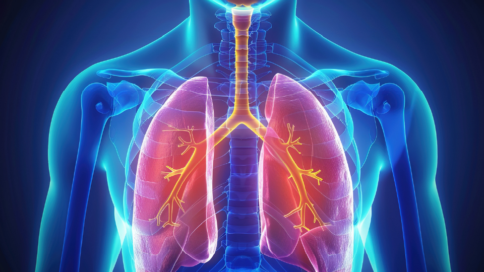
A 78-year-old man with a history of stage 3A colon adenocarcinoma presented with shortness of breath and hypoxia, requiring home oxygen. He previously underwent a curative partial colectomy and later experienced acute illness, progressing to dyspnea, weakness, and an erythematous rash after antibiotic treatment.
Ratings
6
No. of Modules
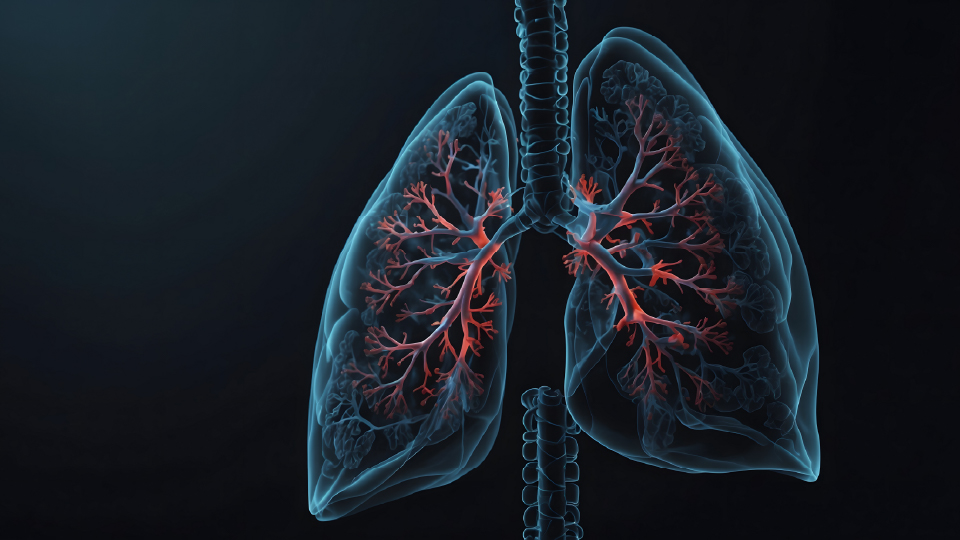
A 58-year-old male experienced an episode of pneumonia five months ago that required treatment, leading to a three-week recovery period before regaining his functional status. Since then, he has developed progressive dyspnea with moderate-to-severe exertion and a persistent, mild, non-productive cough. He denies systemic symptoms such as joint pain, neurological deficits, or edema. His family history is notable for pulmonary fibrosis affecting both his mother and maternal grandfather, as well as his brother, who underwent a bilateral lung transplant for idiopathic pulmonary fibrosis (IPF). Environmental and occupational exposures are minimal-he works as a lawyer, has no smoking or significant allergen exposure history, and lives in a recently built home.
Ratings
5
No. of Modules
A 45-year-old woman with a history of hypertension and mild, persistent asthma presents with progressive dyspnea and dry cough over the course of 9 months. She denies fevers, chills, night sweats, or chest pain. She reported an unintentional weight loss of 10 lb (4.54 kg) and periodic small-volume hemoptysis and epistaxis. She has a 30-pack-year smoking history. She has no travel history and denies any history of substance abuse. She and her husband own and operate a countertop stone manufacturing business.
Ratings
4
No. of Modules

A 53-year-old woman, with a history of asthma on albuterol MDI as needed, presents to the ED with a 3-month history of progressive dyspnea, dry cough, fever, and weight loss. She has a smoking history of 25 pack-years. On examination, her respiratory rate was 25/min and her SpO2 was 90%; on auscultation, bilateral mid inspiratory squeaks and occasional crackles were heard, especially in the middle-to-upper lung zones. A systolic flow murmur was heard as well. Pulmonary function tests, chest CT, and serological evaluations were performed to establish a diagnosis.
Ratings
5
No. of Modules
A 70-year-old male was referred by his primary care physician after a chest X-ray, performed for suspected walking pneumonia, raised concerns about pulmonary fibrosis. He reports experiencing progressive dyspnea with severe exertion over the past 8 to 10 months but remains active. He has a moderate, non-productive cough but denies reflux, joint pain, skin rashes, chest pain, or neurological symptoms, which helps rule out autoimmune causes. His case suggests early-stage interstitial lung disease, warranting further evaluation.
Ratings
5
No. of Modules

A 67-year-old man with no significant medical history was evaluated after an incidental chest computed tomography abnormalities. He was asymptomatic, with normal vital signs and an unremarkable physical examination, but had worked as a pipe fitter and boilermaker for more than 30 years. Pulmonary function testing showed mild diffusion impairment without obstruction or restriction. Bronchoalveolar lavage (BAL) and cryobiopsy demonstrated histiocytic accumulation in the alveolar spaces and interstitium, with minimal fibrosis and preserved alveolar architecture, with Prussian blue staining providing the key diagnostic clue.
Ratings
3
No. of Modules

A 65-year-old man with a 2-year history of idiopathic pulmonary fibrosis (IPF) presented with persistent dry cough despite optimized antifibrotic therapy with nintedanib. The cough had worsened over 6 months, significantly impairing quality of life, and had not responded to nasal corticosteroids or proton pump inhibitor therapy. High-resolution computed tomography showed stable fibrotic changes without evidence of infection or malignancy, and the evaluation excluded gastroesophageal reflux disease, upper airway cough syndrome, smoking, and medication-related causes—raising the question of the next best step in managing refractory cough in stable IPF.
Ratings
3
No. of Modules